Anatomy Yr-1 |
||||||||||||||
 |
||||||||||||
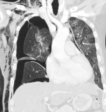
The most anterior cuts of this coronal CT show air dissecting along the fascial planes between muscle bundles of the pectoralis major, accounting for the streaky appearance on the chest radiograph. These are lung window images, but you can still see some soft tissue structures, such as those labeled on Image B.
You can see that the chest tube is going into a space between two lobes of the right lung. What is that space called? Can you tell whether the patient was given IV contrast for this study on these windows?
A
B