Anatomy Yr-1 |
||||||||||||||||
31 year old male with neck pain after motor vehicle collision |
||||||||||||||||
 |
||||||
 |
||||||
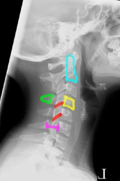
On Image B, the odontoid is shown in aqua, the C3 vertebral body in yellow, facet joints between C3-C4 and between C4-C5 in red, the C4 spinous process in green, and the region where the spinal cord is located is indicated by the purple line segment. |
||||||
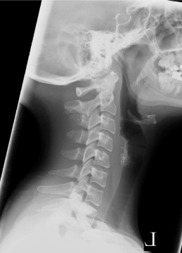
Image A shows the normal lateral radiograph of the cervical spine shown previously. The individual vertebrae are easy to see and the disc spaces show up as dark areas between. This is because discs are made of cartilage, and so show up as low density on radiographs, like the hyaline cartilage of other joint spaces. Image B shows structural elements of the spine for you to identify. |
||
B
A