Anatomy Yr-1 |
||||||||||||||
CASE 2
Patient A
B
C
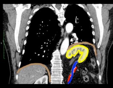
This patient has a congenital diaphragmatic hernia, called a Bochdalek hernia. In this case, it is a very large hernia, with most of the left kidney displaced upward into the chest, stretching the renal artery and vein. The diaphragm on the left is displaced upward. Image B is a select image from Patient A, and Image C shows colored outlines on key structures.