Tufts Anatomy Yr-1 |
||||||||||||||||||||||
 |
||||||||||||||||||||||
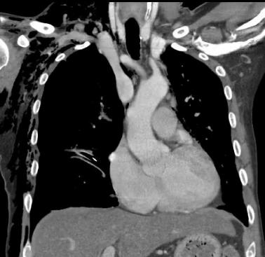
These coronal images are displayed in soft tissue windows. This does not show the lungs or abnormal air well, but does show the structures of the heart and mediastinum. Now it is easy to see that there is IV contrast present. Compare the aorta with arm muscles. The contrast makes the blood in the aorta whiter than muscle. Abbreviations used on Image B:
SCV=subclavian vein BCV=brachiocephalic vein SVC=superior vena cava RV=right ventricle LV=left ventricle RA=right atrium MPA=main pulmonary artery LAA=left atrial appendage (auricle)
A
B
CASE 4 followup