Tufts Anatomy Yr-1 |
|||||||||||||||||
 |
||||||||
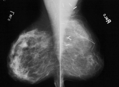
Image A shows a specialized type of x-ray image of the breasts called a mammogram. The view is called an MLO view, or medio-lateral oblique. The beam is coming in from the side at an angle to include part of the axilla. |
||||||||
 |
||||||||
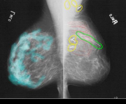
In a mammogram, a specially 'tuned' beam of weak x-rays are used, that show the difference between FAT and SOFT TISSUE most clearly. These are the only tissues normally present in the breast. Because the beam is weak, the radiation dose is low, but the beam cannot penetrate very far, so the breast is imaged in compression, to spread out the tissues and make the breast thinner. In Image B, normal breast soft tissues are shown in aqua on the left, but the right breast has a different appearance. There are surgical clips (metal, circled in yellow) and a wire has been placed on the skin to show the location of a surgical scar (circled in green). There are unusual swirling densities in the upper part of the breast indicated in red, that are not seen on the other side. |
||||||||
B
A