Tufts Anatomy Yr-1 |
||||||||||
 |
||||||
 |
||||||
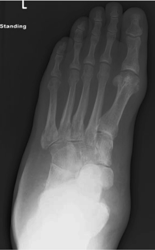
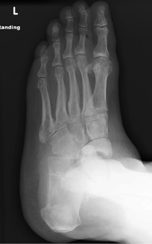
Image A is an AP projection, and there is marked soft tissue swelling in the proximal part of the foot (labeled in red). Image B is an oblique view, at about a 30-40 degree angle, which can show some of the complex joints of the foot better. The joint between the talus and navicular looks abnormally wide (labeled in red), compared to other normal joints (yellow). |
||||||
CASE 1 followup
A
B