Tufts Anatomy Yr-1 |
||||||||||||
65 year old male with right shoulder pain |
||||||||||||
 |
||||||
 |
||||||
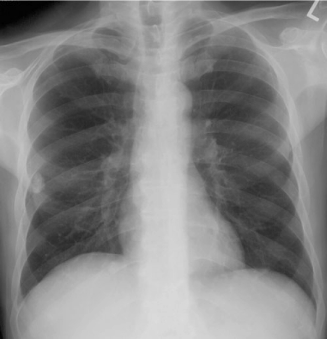
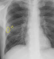
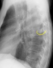
On Images A and C, there is a density (white area) that looks like it is in the outer right lung, about two-thirds of the way down the chest. It is circled in yellow on Image C. If you look closely, you will see that it is also overlapping the lowermost part of the wing of the right scapula. On the lateral view (Image B), it is much harder to see the abnormality, but it is outlined in yellow on Image D. What other type of imaging would be helpful to see this finding better? |
||||||


B
A
D
C
CASE 3 followup