Tufts Anatomy Yr-1 |
||||||||||
43 year old woman with right breast cancer and prior lumpectomy with reconstruction. |
||
 |
||||
 |
||||
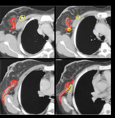
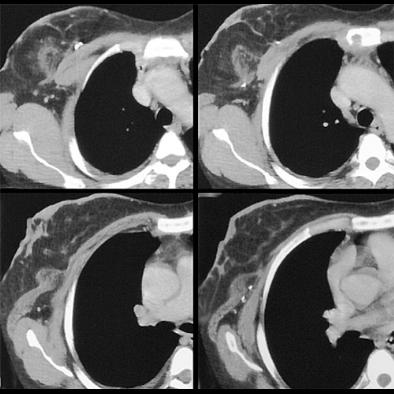
Image A shows additional CT images on our patient, through the region of her breast reconstruction of the right breast. CT is not routinely used to image the breast because of the high radiation dose. Image B shows the swirling muscle fibers (red) and a few of the surgical clips (circled in yellow) that were also seen on the mammogram. |
||
B
A
CASE 2 followup