PrISM Respiratory |
||||||||||||||


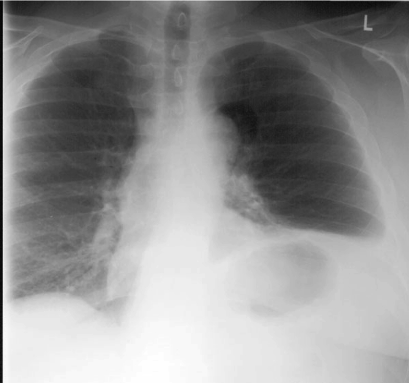
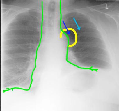
Image A is our patient, and Image B shows labels. The green lines indicate the course of the phrenic nerves, which arise at C3,4,5, travel on the anterior surface of the anterior scalene muscles, and then continue along the lateral margin of the mediastinum on each side, passing anterior to the hilum of the lung, to reach the diaphragms. The abnormality outlined in yellow is best described as a ‘lucency’ or ‘radio-lucency’, and is located along the expected course of the left phrenic nerve, near the level of the aortic arch. How can you relate this abnormality to the prior history of the patient? The dark and light blue arrows indicate that the lucent area is darker than normal adjacent lung tissue.
A
B