Physical Therapy Imaging Cases |
||||||||||||||

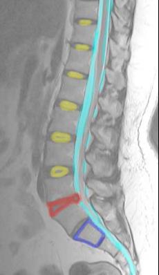
This is a T1-weighted sequence because water is high in signal (white), such as the CSF, which is outlined above in light blue.
There is an abnormal disc at L4-5 (outlined above in red) that is narrowed and lacks the normal central watery high signal of a healthy disc (outlined at other levels in yellow). This is a dessicated disc that has lost height and has herniated slightly posteriorly.
You can tell that this study is an MR right away because FAT is bright (white) and the edges of BONES (cortical bone) are dark (the cortical bone of S1 is outlined above in dark blue). This is the opposite of how fat and cortical bone look on CT.
For comparison, above is the T1-weighted sequence on the same patient. Look at the CSF and you will see that it is now DARK. This is how you recognize a T1-weighted sequence--areas of water or watery fluid (like urine, bile or CSF) are dark on T1-weighted images, and bright on T2-weighted images.
The abnormal disc is only bulging posteriorly a small amount. Is this the cause of the patient's pain? It is almost impossible to tell. MR of the lumbar spine can be problematic for just this reason-it may detect abnormalities that may not have anything to do with the patient's symptoms. This patient improved with conservative treatment and never had any symptoms that specifically localized to the L4-5 level.