Physical Therapy Imaging Cases |
||||||||||||
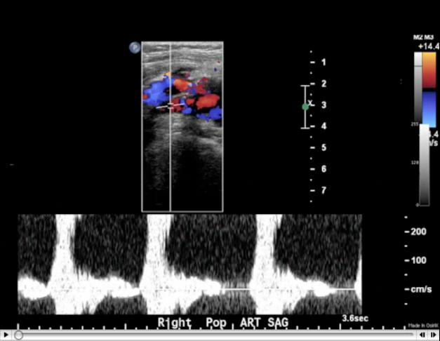
The top image is part of a popliteal ultrasound study, and the colored areas are Doppler flow analysis. The mixture of blue and red areas indicate tubulent flow. The scale on the right-hand side of the image shows that red colors indicate flow TOWARD the transducer and blue colors indicate flow AWAY from the transducer. The transducer is the mechanical apparatus that is used to send sound waves in and record reflected waves bouncing back. In this case, the transducer is touching the back of the patient's knee, in the popliteal fossa, so flow indicated in red is toward the back and blue is toward the front. Red and blue do NOT correlate to arterial and venous flow, just directions. The bottom tracing shows an arterial pulse in the area of the abnormality, indicating that it is a popliteal aneurysm.
You have finished the lower extremity images, and can go on the the next topic.
