PA Anatomy: Imaging Overview- MR |
||||||
 |
|||||||||
On routine T1 and T2-weighted MR images fat is high signal (white). In some parts of the body, this can interfere with detection of other abnormalities. So additional sequences can be performed, in either T1 or T2-weighted mode, with what is called 'fat suppression' or 'fat saturation'. This decreases the signal from fat, so that fat becomes dark, similar to the appearance on CT. This is why it is not always sufficient to look at fat to determine whether an image is a CT or an MR. If fat is white, you KNOW it is in MR. But if fat is dark, it could either be a CT or an MR with fat suppression. |
|||||||||
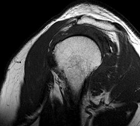
This is a sagittal image of the shoulder, with T1-weighting. There is actually a small joint effusion (fluid in the joint) but it is very hard to see because it is low signal and is adjacent to the low signal of the bone. |
|||||||||
 |
|||||||||
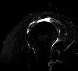
This is a sagittal image of the same patient, performed with T2-weighting and fat suppression. The only high signal areas are areas of fluid, which really stand out and show the joint effusion very clearly. However, the rest of the anatomy of the shoulder is hard to see without bright fat to outline the muscles.. So you do need to do both imaging sequences and look at them together to be able to figure out what is going on. |
|||||||||