PA Anatomy: Imaging Overview- CT |
||||||
As previously noted, for use in the vascular system, only Iodine-based contrast is appropriate. For CT, just like with oral contrast, whether to use IV (intravenous) contrast depends on the particular clinical setting. And just like for GI contrast, timing is crucial. IV contrast is usually injected into an arm vein, so it will first appear in the upper body veins, then in the arteries to the lungs, then in the arteries going out to the body via the aorta (including kidneys), and last in the lower body veins. Scanning should begin at different times after injection, to maximize visualization of specific vessels and organs. |
||||||||||||||
 |
||||||||||||||
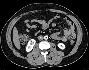
Early IV contrast-aorta (A) is very white, much whiter than muscle (M), and kidneys (K) are also very white around their edges, with darker grey centrally. The major lower body vein, the inferior vena cava (V) is not yet white, and is the same color as muscle. |
||||||||||||||
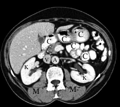
Later after IV contrast injection-aorta (A) and inferior vena cava (V) are now similar in color, both lighter than muscle (M). Kidneys (K) are uniformly white. The amount of time it takes for IV contrast to circulate around from arteries to veins depends on cardiac function. Note that there is also oral contrast (C) in GI structures. |
||||||||||||||
 |
||||||||||||||
 |
||||||||||||||
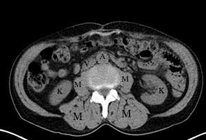
CT image without oral or IV contrast-the only white areas are bones. The aorta (A), inferior vena cava (V), kidneys (K) and muscles (M) are all the same shade of grey. Blood is similar in density to muscle. |
||||||||||||||