PA Anatomy: Imaging Overview-Contrast |
||||||
 |
||||||||||||
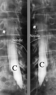
This is a study called a 'myelogram', in which iodine-based contrast is injected through a needle inserted into the lower back into the thecal sac, or the lining that surrounds the spinal cord and nerve roots. These are two oblique views showing the contrast in the lower lumbar and sacral regions (C). The needle is visible overlying one of the lower lumbar vertebrae. |
||||||||||||
Barium cannot be injected into body cavities or the vascular system. Iodine-based (water-soluable) contrast must be used for these types of studies. Iodine-based contrast material can induce a mild or severe allergic reaction in some people, and cannot be given to patients who have poor renal function, as it can damage their kidneys. |
||||||||||||
 |
||||||||||||
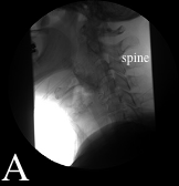
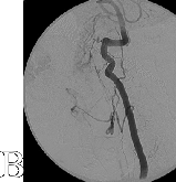
This is an example of an 'angiogram', which is a general term for injection of iodine-based contrast material into some part of the cardiovascular system. If it is injected into a vein, it can also be called a 'venogram', and if it is injected into an artery, it can be called an 'arteriogram'. This study is of an important artery in the neck, the vertebral artery, so it can be called a 'vertebral arteriogram'. This also shows that if you start with a radiograph before injecting contrast (image A, a lateral view of the neck, with the spine to the right), and the patient does not move, then you can subtract the background from all subsequent images so that only the contrast is visible (Image B). This is called 'digital subtraction', and makes tiny vascular structures easier to see. |
||||||||||||
 |
||||||||||||