PA Anatomy: Brain: Case 1 |
||||||
 |
||||
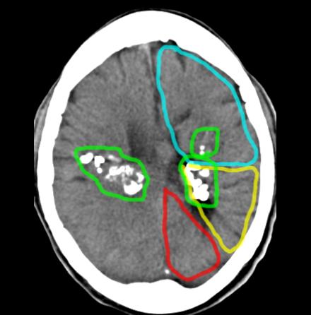
The parietal lobes are most involved bilaterally, but there are many other areas of involvement. Frontal lobe is shown in blue, parietal in yellow, and occipital in red. The abnormal areas of high density are outlined in green and represent calcifications. |
||||
There is no IV contrast on this study. The next image shows a comparison of a normal case, with and without IV contrast to help you figure out how to tell them apart. |
||