PA Anatomy: Pelvis: Case 4 |
![]() |
|||||
![]() |
||||||
![]() |
![]() |
![]() |
![]() |
![]() |
![]() |
![]() |
![]() |
||||||||||||
![]() |
![]() |
![]() |
![]() |
![]() |
||||||||
![]() |
![]() |
![]() |
![]() |
![]() |
![]() |
![]() |
![]() |
![]() |
![]() |
![]() |
![]() |
![]() |
![]() |
![]() |
![]() |
![]() |
|||||
![]() |
![]() |
![]() |
![]() |
![]() |
![]() |
![]() |
![]() |
![]() |
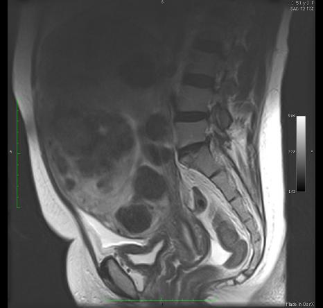
There is watery fluid in several regions on this image, indicated in yellow. The best place to look is in the bladder. There is also a small amount of water in the cervical canal and in the vagina. If WATER is high signal (white), then the image is T2-weighted! T2-weighted images often show pathology best. |
![]() |
|||
![]() |
![]() |
![]() |
![]() |
![]() |
![]() |
![]() |
![]() |
![]() |
|||||
![]() |
![]() |
![]() |
![]() |
![]() |
![]() |
![]() |
![]() |
![]() |
![]() |
![]() |
|||
![]() |
![]() |
![]() |
![]() |
![]() |
![]() |
||||||
What do you think of the uterus? |
![]() |
|||||
![]() |
![]() |
![]() |
![]() |
![]() |
![]() |
![]() |