PA Anatomy: Abd: Case 1 |
||||||
 |
||
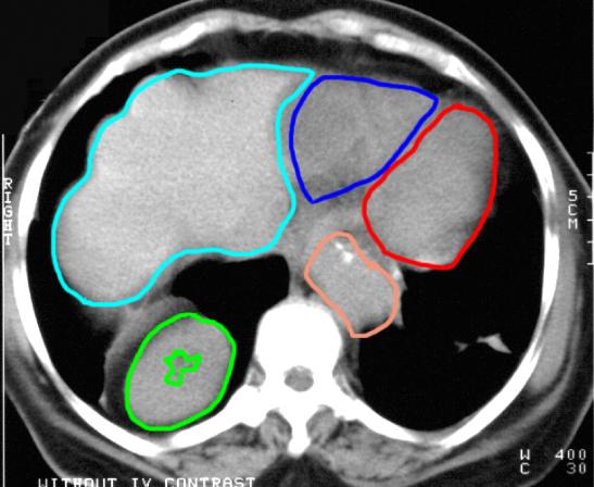
This is a CT scan (bones are very white, fat is very dark), in the axial plane, no IV contrast, soft tissue windows (muscle and fat look very different in color). Try to determine what each of the labeled structures are on this image. The mass that was seen on the chest radiograph is outlined in green. What organ could this be? |
||