Anatomy: Imaging Overview- US |
||||||
 |
||||||||
One of the limitations of ultrasound is that the field of view (portion of the body represented on the image) is quite small. You cannot get an image of a whole slice of the abdomen at once, like you can with CT and MR. It is like you are looking at the body through a narrow window. US beams are also blocked by certain relatively common tissues or substances, in particular BONE and AIR. So US is not used in imaging the lung, or deep bony structures, although it can be used to image tendons and ligaments around bones if they are superficial in location. |
||||||||
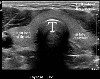
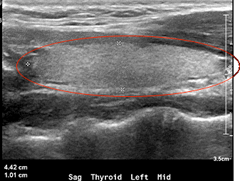
Because the amount of detail that is visible in US images is limited, most images have extensive labeling that is entered by the technologist or radiologist who is doing the study (writing at the bottom of each image). These two images are of the thyroid gland in the neck. 'TRV' means transverse, or axial. 'Sag' means sagittal. These are slices, just like CT or MR. There is a black band extending from the front of the trachea (T) to the bottom of the image.. That is because the air in the trachea completely blocks the beam, so no information can be obtained deep to the tracheal air column. |
||||||||
 |
||||||||
Red circle shows the long axis of the left lobe of the thyroid, which is being measured |
||||||