Anatomy |
||
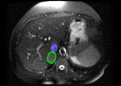
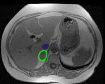
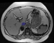
Image A is a T2 weighted image and Image B is a T1-weighted image, both showing a rounded right adrenal mass, just posterior to the IVC. Image C is a special type of MR image for characterizing adrenal masses. The signal in the adrenal mass will become quite low on this sequence if the mass contains intracellular fat, which is typical of a benign adenoma, which can produce excess secretions leading to Cushings syndrome, as in this case. |
||


A
B
C