Anatomy |
||||
 |
||||
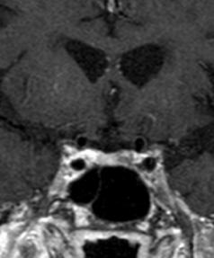
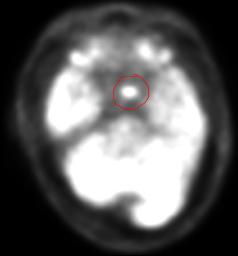
This patient has a focal area of increased uptake on the PET scan, Image A (done by injecting a radioactive analog of glucose intravenously, and waiting for uptake by normal brain and by metabolically active regions of the body, often tumors, either benign or malignant). The abnormality location suggests the region of the pituitary gland, and an MR is shown in Image B with a small tumor in this same area, an adenoma. Normal pituitary is shown in green, and adjacent carotid arteries in red. The next cases in this module will explore imaging of the pituitary in more detail. Even such a small adenoma can secrete enough ACTH to lead to adrenal hyperplasia, as in this case.

A
B