Anatomy |
||||||||||||||||||||
 |
 |
|||
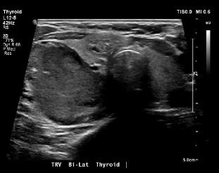
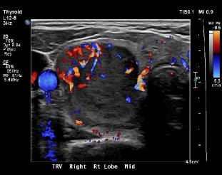
On each labeled image, the mass is in red and the normal thyroid in yellow. When looking at thyroid US, it is often helpful to find the trachea, labeled in blue on Image A. Be sure to look at the writing on each image to determine if you are looking at a sagittal or a transverse view. Remember, the transverse view is displayed like an axial CT or MR image, with the patient's right on the left. The sagittal view is shown with the patient's head to the left of the image. Two features that are important in analyzing thyroid nodules are SIZE and ECHOGENICITY. Nodules that are over 1 cm, and nodules that are hypoechoic (darker) than surrounding thyroid tissue are more worrisome. Increased vascularity is also somewhat concerning. This nodule is well over 1 cm, hypoechoic, and very vascular. |
||
A
B