Anatomy |
||||||||||||||||||||


A
B
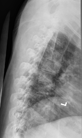
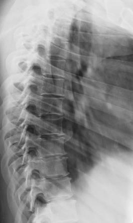
Image A shows our patient's lateral thoracic spine radiograph, performed for back pain. The spine is very washed-out in appearance and hard to see due to marked osteopenia. A normal comparison in a different patient is shown in Image B, where each vertebra and disc space can be clearly seen. Radiography cannot quantify the degree of bone loss, but can detect overall decreased density as well as comlications, like compression fractures. For quantification, bone densitometry is indicated.