Anatomy |
||||||||||||||||||||
 |
||||
 |
||||
 |
||
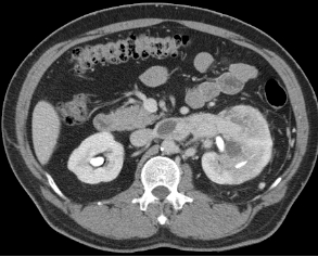
Image A shows a large mass replacing the mid and lower pole of the left kidney and invading into the left renal vein, a pattern of spread that is typical of renal cell carcinoma.
A
B
C
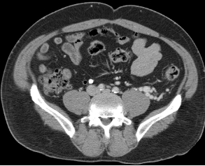
Image B shows many dilated vessels just anterior to the left psoas muscle. Recall that the gonadal veins drain not into pelvic veins but into the IVC at the level of the kidneys.
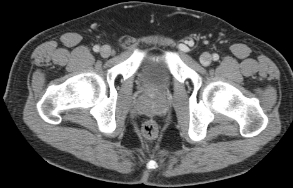
Image C shows many dilated vessels in the left spermatic cord (muscle layer of both spermatic cords outlined in orange). These vessels represent a varicocele due to obstruction of venous return at the level of the kidneys from the large tumor.