Anatomy |
||||||||||||||||||||
 |
||||
 |
||||
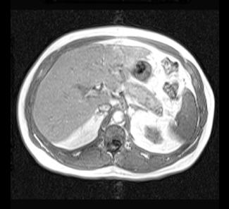
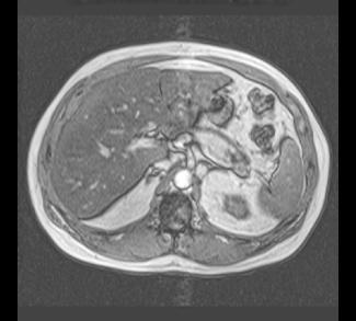
These are both MR images (note the dark appearance of bone and high signal fat). The image on the left is an in-phase image and the image on the right is an out-of-phase image, which causes a number of artifacts (a black outline around many structures, called 'India Ink' appearance) but also causes signal from fat to decrease. The liver is brighter on the in-phase than the out-of-phase image, indicating that it has diffuse fatty infiltration. MR is not used as frequently as CT overall in abdominal imaging, but can solve particular problems, such as detecting small amounts of fat within organs or lesions, which can often help in the differential diagnosis of complex cases.