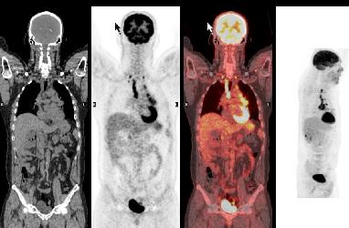
Anatomy |
||||||||||||||||||||
 |
||||||||
 |
||||||||
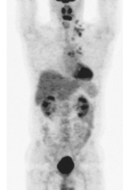
Normal areas of uptake on this PET scan are shown in aqua, and abnormal areas in red on the labeled image.
This is a study on a different patient. What is this study? Where are normal and abnormal areas of uptake? (check by putting labels on)
PET stands for Positron Emission Tomography, and is a type of nuclear medicine study, meaning that a specially designed radio-tracer is introduced into the body to show either a normal or abnormal physiological process. In the case of PET, the agent shows where glucose is being metabolized, which happens in some normal tissues as well as in many tumors. So we expect to see uptake in the brain, myocardium and renal system, and to a lesser degree in liver and spleen.