Anatomy |
||||||||||||||||||||
 |
||||||
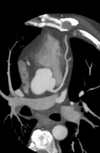
The data from the CTA on the left can be reconstructed into a 3D representation, as shown on the previous page. The movie on this page has had cardiac structures made translucent to better demonstrate the normal anatomy of just the aorta and coronary vessels. Faster scanning and better computer algorithms for reconstruction have made CTA a more valuable method for evaluation of coronaries than in the past, and is less invasive than angiography. |
||||
What is this study called and what is the imaging plane? Is it normal? This is a CT of the coronaries, or CTA (CT arteriogram). This requires administration of IV contrast and careful timing so that the majority of the contrast is in the aorta at the time of scanning. This image is in the axial plane. Click to see labels of the coronary structures on this slice. This is a normal study. |
||||