|
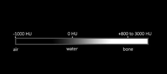
Because there are so many different tissue densities that can be displayed with CT, we often use specific brightness and contrast settings (called "CT windows") to optimize viewing of particular regions. For the lung, we use most of our display shades of gray in the negative end of the density spectrum, which produces an image that shows detail in the lung, but most of the denser tissues (like muscle or organs or bone) look uniformly white. It is important when viewing CT images to be sure you are using the right window for the abnormality you are evaluating. If you look at the previous CT image, you will see the lung well, but the chest wall and other soft tissues show no details.
|
|

