|
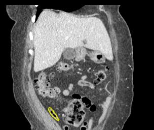
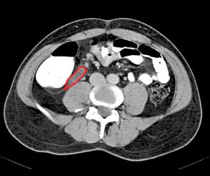
Which one has a normal appendix? The patient on the left has a dilated appendix filled with fluid (red), and a little bit of stranding in the surrounding fat. This is not normal and indicates appendicitis. The patient on the right has a normal thin appendix containing gas (yellow) on this coronal reconstructed image. This rules out appendicitis. If the appendix is NOT visualized (as in our patient), then appendicitis is still a possibility. Sometimes a normal appendix may be hard to see if it is positioned to close to other structures, or it might be hidden by surrounding fat stranding.
|
|