|
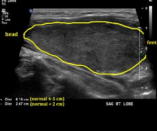
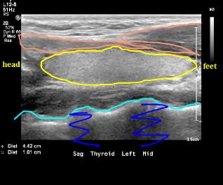
What is the orientation of these two images? These images are in the sagittal plane (look for a label that says 'Sag'), so the sonographer turned the transducer 90 degrees. By convention, when displaying sagittal images, the patient's head is to the left of the image and their feet to the right. So the upper pole of the examined thyroid lobe is to the left of each image and the lower pole is to the right. Ultrasound is great for accurately measuring the size of structures. The measurements are shown on the images. The patient on the left has a right lobe that measures over 6 cm in superior-inferior length (normal is less than 5) and 2.5 cm in AP dimension (normal is less than 2). On the normal comparison, you can see some neck muscles (orange) and the front surface of the cervical spine (aqua) with shadowing behind (dark blue).
|
|