|
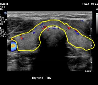
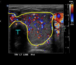
What do these two images show, in terms of technique? These are both color flow images, with flow toward the transducer shown in red and away shown in blue (there is a color scale on the image on the left to show this range). What do you think the significance is of the different appearance of the two studies? There is markedly more flow throughout the thyroid in the patient on the left, suggesting an overall stimulated metabolic state of the whole gland. The gland is so large that the transducer has been shifted to show just the left lobe of the thyroid, with the trachea off to the other side (letter T). This increased flow is typical of thyroiditis, including Graves disease, but must be correlated with results of thyroid function testing.
|
|


