|
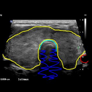
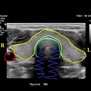
Patient study is on the left, normal comparison on the right. See if you can identify the thyroid gland. The thyroid gland is outlined in yellow. How does it look different comparing the two examples? The patient study on the left shows a large and slightly lumpy gland with thickening of the isthmus, the part of the gland that passes in front of the trachea (aqua) and connects the right and left lobes. What is the imaging plane and which side is the patient's right side? These images are in the transverse plane (label on the right says 'TRV') and as for CT and MR images, the patient's right is on the left side of the image, as if you are looking at them from their feet (like you would when coming into a room to talk to a patient who is lying on an exam table). The carotid artery is outlined in red. Shadowing behind the air in the trachea is indicated in dark blue.
|
|


