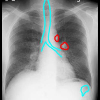
ICM-II: CHEST, case 1 |
||
TERMINOLOGY-opacity/ lucency
It is important to use the right terminology for each type of imaging that is used. For radiography (no matter what body part), we call white areas opacities or densities, and dark areas lucencies. |
||||||
 |
||||||
For example, on this image there are several areas that appear WHITER than surrounding tissues (outlined in red), and should be called 'densities' or 'opacities'. The lungs are more LUCENT than all surrounding tissues, as is the trachea and air in the colon (outlined in blue). The actual degree of whiteness or darkness of a particular pixel on the image is a sum of all the different tissue densities encountered by that particular portion of the x-ray beam on its way to the receptor. Can you identify what the DENSEST area on this image is? |
||||||
 |
|||||||||||