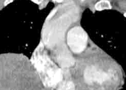
normal CT for comparison
Here is a selected image from the normal study. This study is not as well-timed as the other patient's CTPA, with less concentrated contrast within the pulmonary arteries. The main pulmonary artery is shown in purple, the ascending aorta in red, the right atrium in dark blue, the right ventricle in light blue, and the left ventricle in green. The images are displayed in the coronal plane.

