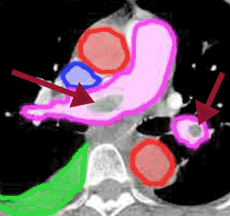
Case 6: shortness of breath due to pulmonary embolism

This study is a CT angiogram of the pulmonary arteries. A selected image from the study is shown here, with and without labels. The pulmonary arteries are outlined in purple. The aorta is outlined in red. The superior vena cava is outlined in blue. A small right pleural effusion is outlined in green. Notice that the contrast is brightest in the SVC, indicating that the scan began soon after IV contrast was administered (in an arm vein). Notice also that the pulmonary arteries have much more contrast than the aorta, another indication of early scanning. The timing for this study is vital to detection of the emboli, which can be seen in the right and left pulmonary arteries on these images (dark red arrows). In ordinary contrast-enhanced chest CT scans, the scanning is done at a later point after contrast administration, when the contrast is more equalized between aorta and pulmonary vessels, an equilibrium phase. With less concentrated contrast in the pulmonary arteries, small emboli may be missed. Large emboli like these would probably still be visible even on a scan done at equilibrium.
