Case 5: SOB due to diffuse interstitial metastases from breast cancer

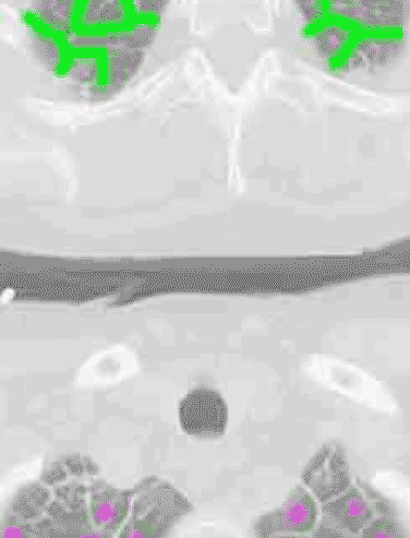
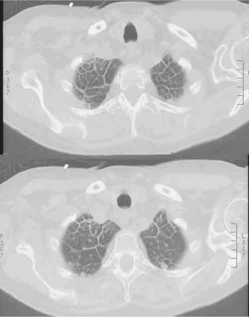
On the CT, you can see little polygonal shapes bordered by thin white lines (indicated in green). This is the tumor growing along the supporting struts of the lung, the interstitium, which is made up of connective tissue. Just like elsewhere in the body, tumor often grows along fascial or connective tissue planes. When this happens in the lungs, it produces a reticular pattern on chest radiographs of criss-crossing lines as seen in the original chest radiograph of this case. In the center of each polygon is a little dot, shown here in purple. These dots are the smallest pulmonary artery branches that we can see on CT and are normally all that is visible in this part of the lung. There is a bronchus running with each, but they are too small to see. The pulmonary veins run along the connective tissue struts.

