typical effusions
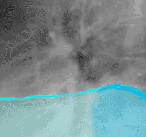
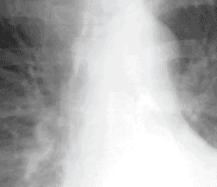
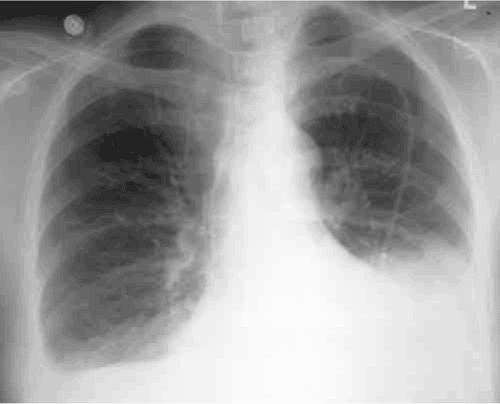
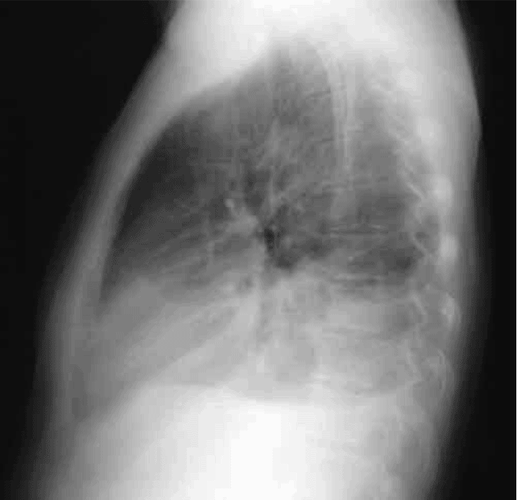
This patient has bilateral pleural effusions, larger on the left than the right. The meniscus (arrows) shows the fluid creeping up the inside of the ribcage due to surface tension, producing the characteristic “blunting” of the costophrenic angles. In addition to the relatively sharply defined margin of fluid against lung (shown in light blue on the left and light green on the right), there is also a more hazy opacity that is due to fluid in a different plane (shown in darker blue and darker green). REMEMBER that ‘on the right’ refers to the patient’s right, which is on the left side of the image, since we view frontal radiographs as if we are facing the patient. As shown on the lateral view from this same patient, the interface of lung to pleural fluid occurs at different levels as you move from anterior to posterior. Both effusions also produce posterior menisci on this view. The larger left effusion is shown in blue and the smaller right effusion in green.


