Case 1: shortness of breath due to pleural disease (mesothelioma)

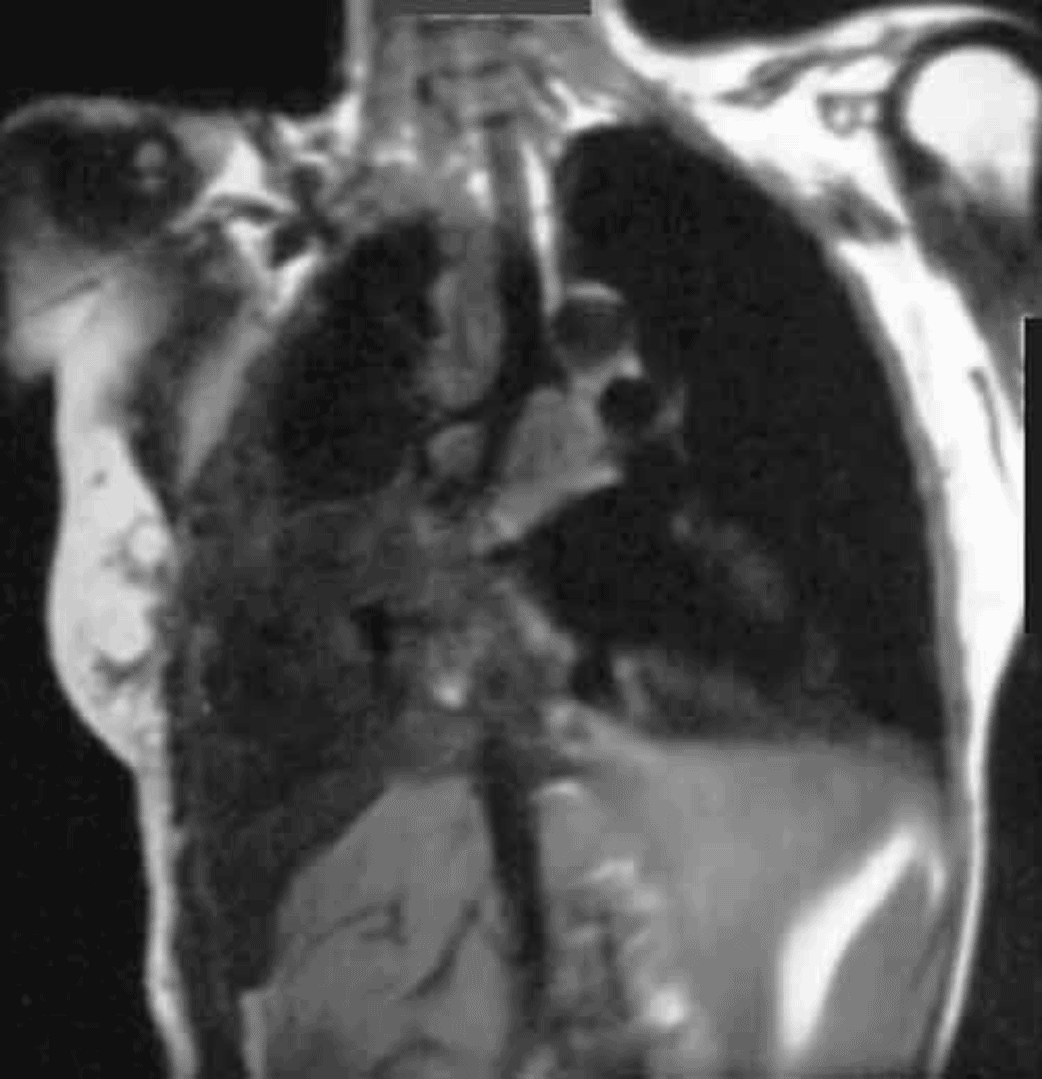
This is a chest MRI in the coronal plane. The coronal plane cuts the body into anterior and posterior portions. You can tell that this is an MRI because the fat (outlined in blue) is bright, or high in signal. Fat on CT is dark, or low in attenuation. The bones also appear dark on MR, other than the fatty marrow, seen here in the humeral head. The mesothelioma is outlined in red, and is filling much of the right hemithorax.

