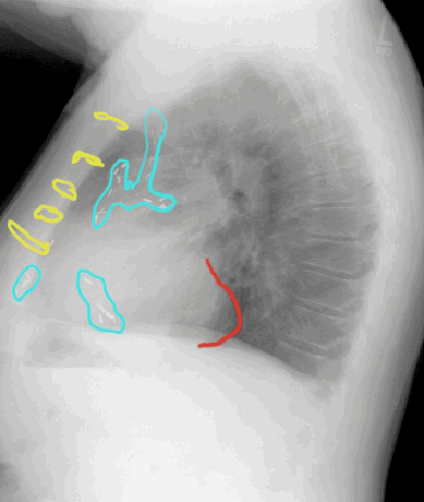
The sternal wires are shown in yellow and the surgical clips are outlined in blue. There is a focal out-pouching or bulge along the posterior border of the heart (red). Is this the same location as the ventricular aneurysm shown in the previous case? What other imaging could you do to better define this abnormality?
Lateral radiograph, labeled
labels off
