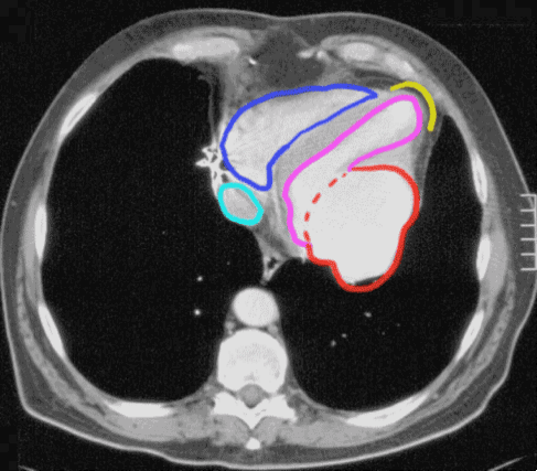
On this single CT image, the right ventricle is shown in dark blue, the IVC in light blue, the left ventricle in purple and the usual location of an aneurysm of the left ventricle in yellow. The out-pouching of the left ventricle in this patient is shown in red, and is protruding posteriorly, rather than at the apex. This is typical of a pseudoaneurysm or contained ventricular rupture, which requires immediate surgical repair.
Cardiac CT, labeled
labels off
