CT of Collapse
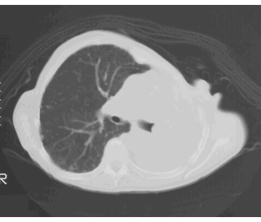
CT image from the patient with breast cancer and collapse. The left lung is totally opaque and very small, while the right lung extends past the midline to fill in the space.
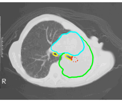
On this labeled image, the heart is shown in blue, markedly shifted to the left. The two mainstem bronchi are shown in yellow and the mass in red. The entire mass is not visible, just the end that abuts the air-filled bronchus, producing a contour called a 'capping defect'. The opaque left lung is outlined in green. When analyzing cases with altered lung VOLUME, it is important to look for secondary signs, like shift of the mediastinum, elevation or depression of the diaphragm, and changes in the spacing of ribs. How would you expect spacing of ribs to differ in the right vs the left in this patient?


