Two other patients with wheezing and fever


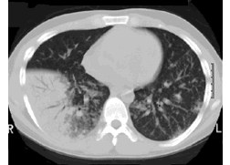
A
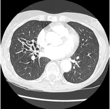
B


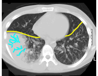
This patient has abnormal AIRSPACES, which make his normal bronchi (blue) stand out. The problem is in the tissues surrounding the bronchi, not in the bronchi themselves. The bronchi are normal but just better seen due to the dense opacity in surrounding lung. Normal lung is seen anterior to both major fissures (yellow). These black branching structures running through a background of abnormally white lung are called AIR-BRONCHOGRAMS, in this case due to infiltration of the lung by lymphoma.
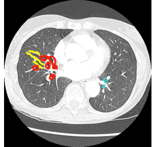
This patient has abnormal BRONCHI on the right, which are irregularly dilated with thickened walls. When these thickened walls are seen in cross-section, they appear as rings (peribronchial cuffing, shown in red). When they are seen in longitudinal section, they appear as irregular parallel lines (tram tracking, shown in yellow). Normal bronchi for comparison on the left are shown in blue. The right findings are called bronchiectasis, and in this case were from old pneumonia in this region.
