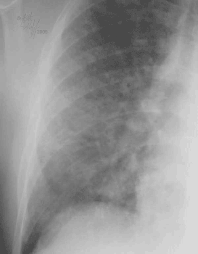
Set 3: air bronchograms

Here is the image with labels showing the small branching tubular structures that can be seen in the areas of most dense alveolar opacity. These are called ‘air-bronchograms’. The bronchi themselves are not abnormal. They just become visible because the lung all around them has become filled, in this case with acute edema fluid from CHF. In general, more chronic forms of CHF will have fluid in the interstitium (sometimes with Kerley B lines) and more acute or severe forms will have fluid in the alveoli (sometimes with air bronchograms, as in this case).
