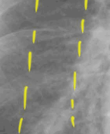
Set 2: interstitial edema
lung zones

It is helpful to divide the lung into three zones for analysis of interstitial processes and CHF, as shown here in our original patient. In the inner zone, there are many large overlapping vessels. In the middle zone, you should be able to trace out vascular branches. In the outer zone, there should be almost NO lung markings. In this case, the inner and middle zone vessel margins are not very sharp, and there are many lines in the outer zone, including some horizontal Kerley B lines.

