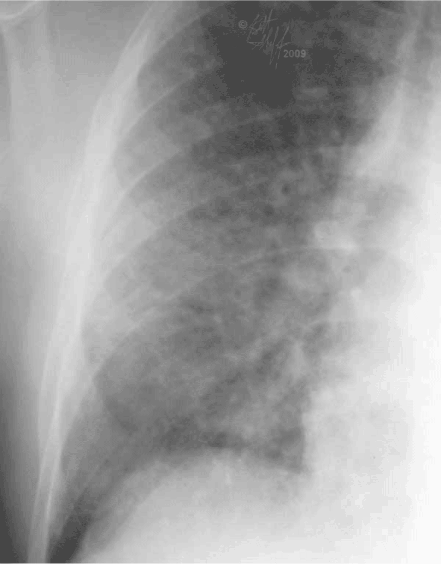
Set 1: interstitial vs alveolar
45 year old male diagnosed with a new large myocardial infarction

In this case, the abnormal lung opacities are much less well-defined, looking fluffy like cumulus clouds. This is an example of an ALVEOLAR pattern. Alveolar abnormalities tend to look out-of-focus or ill-defined. Both interstital and alveolar patterns can often be caused by the same conditions, such as lung edema or hemorrhage, so they are NOT specific to a particular diagnosis. In this case, because of the history of recent heart damage, this is likely due to pulmonary edema.

